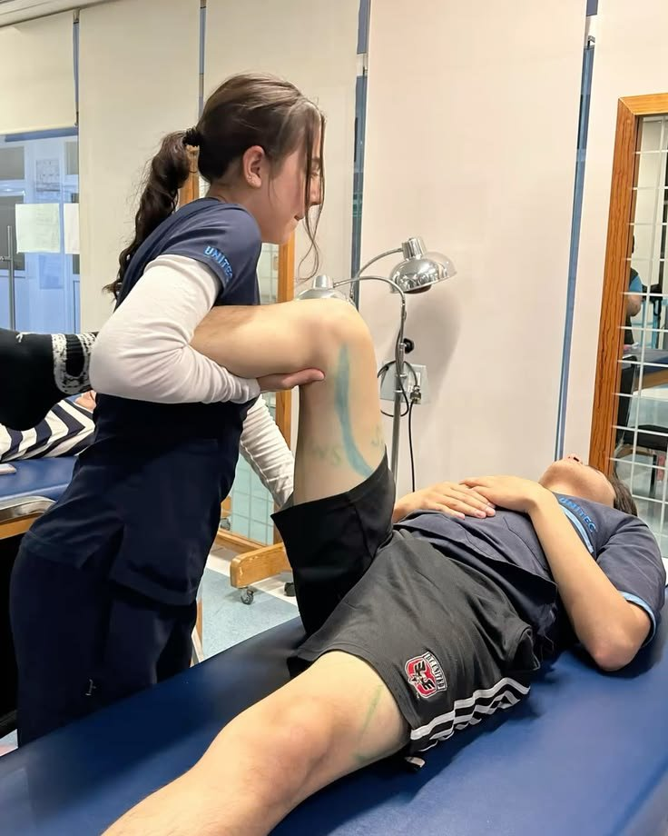
After PRP for a chronic tendon problem, a repeat MRI is not automatically the best way to decide whether treatment has worked. Pain, function, examination and the ability to tolerate the activities that matter to you need to be reviewed together. A scan can answer a specific clinical question, but an image is not a standalone clearance certificate for running, lifting or returning to demanding work. [1]
Ask The London PRP Clinic about the review plan before treatment. Agree what will be measured, who will review it and what would lead to further imaging. This guide concerns chronic tendinopathy after an injection, not acute tendon rupture or postoperative restrictions.
What question would the repeat scan answer?
Before booking another scan, ask the clinician what uncertainty it is intended to settle. Is there a suspected new tear, an unexpected change in symptoms or a diagnosis that no longer fits the examination? Those are different questions from wanting a picture that proves healing. The appropriate investigation depends on the clinical question and the tendon involved.
A 2026 international expert consensus on rehabilitation after PRP for chronic tendinopathy supported basing return-to-activity decisions on clinical assessment rather than requiring imaging. That does not mean scans are never useful. It means a routine image should not replace the assessment of how a person actually functions. The authors also emphasised that many recommendations rested on low-level evidence and expert opinion. [1]
Measure the activity you want back
Describe the task that was difficult before treatment and what happens now. For one person that may be walking downstairs; for another it may be lifting at work, playing tennis or running for a sustained period. Keep the task consistent enough to make changes meaningful, while following the loading limits set by your clinician or physiotherapist.
A review can consider pain during activity, the response afterwards, strength, confidence and the amount of activity tolerated. The consensus supported including a pain-and-function score, such as a tendon-specific questionnaire, although agreement and evidence strength varied across recommendations. A score is a structured aid to the conversation, not a diagnosis or proof that PRP caused an improvement. [1]
Why a better-looking image is not the whole outcome
Research imaging can measure changes that routine scan descriptions do not capture. A small randomised patellar tendinopathy trial compared PRP, needle tenotomy and sham injection in 29 participants, measuring clinical outcomes alongside ultrasound and specialised MRI markers. All groups improved in pain at 52 weeks, with the largest improvement in the PRP group. Some imaging measures changed or correlated with pain, but the authors called for further research into their clinical applicability. [2]
That finding is interesting, not a mandate to order advanced imaging for every tendon. The sample was small, most participants were men and the findings concerned patellar tendinopathy. They cannot establish that every tendon needs the same scan, that a particular image proves recovery or that PRP will produce the same result in another diagnosis. A useful outcome remains one that matters to the patient as well as the investigator.
A follow-up is not automatically another injection
At review, discuss whether symptoms are improving, unchanged or worse and whether the rehabilitation plan has been possible to follow. A partial response should prompt a conversation about the diagnosis, loading and next step, not an automatic sale of more treatment. The 2026 consensus suggested that a second injection could be considered after three months in selected partial responders, but this was an expert-consensus recommendation with low-level evidence, not a universal schedule. [1]
Do not use that interval as a reason to wait through a new injury or concerning deterioration. Equally, do not assume that an unchanged scan requires another injection when function is improving. Ask the clinician to explain what additional treatment is intended to achieve, the evidence for your tendon, its cost and what would change the recommendation.
Who should decide when activity progresses?
Your treating clinician and rehabilitation professional should agree how activity progresses, accounting for the diagnosis and your demands. The consensus supported a final rehabilitation phase that includes exercises related to the patient's work or sport. A short painless walk does not recreate a shift involving heavy lifting or a long run. [1]
Make the handover explicit when different professionals are involved. Ask who will review progress, which symptoms should trigger contact and whether the physiotherapist has the procedure details and restrictions. A general article, scan report or package description cannot safely replace individual clearance. Different tendons, associated injuries and postoperative plans may require different rules.
What to agree before paying for follow-up
Ask what the review includes, whether a scan is proposed and why, whether the images will be compared with a useful baseline, and what the next decision will be if symptoms and imaging disagree. Confirm fees and any separate imaging costs directly rather than assuming they are included in a treatment package.
Review services and ask for an individual follow-up plan. Bring the original diagnosis, procedure date, rehabilitation record and prior imaging reports. The aim is a clear clinical decision, not simply another photograph of the tendon.
For information only. Do not change activity restrictions or medicines on the basis of this article. Sources checked November 2026.
References
[1] Gremeaux and colleagues, Rehabilitation and return to activity after platelet-rich plasma injections in chronic tendinopathies: Consensus from international experts, PM&R, 2026. Expert consensus; important evidence limitations apply.
[2] Platelet-rich plasma for patellar tendinopathy: a randomized controlled trial correlating clinical outcomes and quantitative imaging, PubMed record. Small trial of 29 participants; not evidence for routine imaging across all tendons.