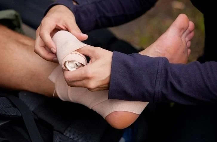
Ankle support is one part of a broader assessment and rehabilitation plan. Front-of-ankle pain needs a diagnosis before an injection is considered. Illustrative photograph.
A pinching pain at the front of the ankle can make a deep squat, an uphill walk or a return to running unexpectedly difficult. Sometimes it follows an ankle sprain. Sometimes it develops gradually with repeated loading. Anterior ankle impingement describes a mechanical restriction involving bone or soft tissue at the front of the joint, rather than a single injury with one standard treatment.
The distinction matters when considering platelet-rich plasma, or PRP. An injection cannot be assumed to remove a bone spur or correct an unstable ankle. A useful assessment establishes what is being pinched, whether other joint damage is present and which part of the problem a proposed treatment could reasonably address. Clinical review comes before choosing an injection.
What does anterior ankle impingement mean?
The ankle bends upwards when the shin moves towards the foot. This movement is called dorsiflexion. During a squat, an uphill step or a forward lunge, the structures at the front of the ankle move closer together. If an abnormal bony prominence or thickened soft tissue occupies that space, the movement can become painful or restricted.
The British Orthopaedic Foot and Ankle Society describes ankle impingement as a painful mechanical limitation of movement caused by a bone or soft-tissue abnormality. At the front of the ankle, repeated loading can contribute to inflammation of the joint lining, while bony changes can create a more structural block. These mechanisms can coexist rather than fitting neatly into separate categories.
The name does not establish the cause by itself. Front-of-ankle pain can also occur with arthritis, cartilage injury, tendon problems and other conditions. A symptom that feels like a pinch is a reason to examine the joint, not a reliable way to diagnose impingement without an assessment.
Why can it follow an ankle sprain?
An ankle sprain can leave more than temporary swelling. Scar tissue, persistent joint-lining inflammation and changes in how the ankle moves can all become relevant to ongoing symptoms. Some ankles also remain unstable after ligament injury. A feeling that the ankle gives way may therefore be important alongside the location of pain.
BOFAS describes an association between anterior impingement and chronic ankle instability. A deficient anterior talofibular ligament can alter the position of the talus during movement. Thickened ligament tissue and post-traumatic fibrous bands are other possible contributors. These are clinical explanations to consider, not a conclusion that every old sprain has left permanent damage.
The assessment should connect the current symptoms with the original injury and recovery. Persistent pain despite rehabilitation may reflect an untreated contributor, a different diagnosis or a rehabilitation plan that needs adjustment. Repeatedly treating the painful area without reviewing stability can miss part of the reason symptoms continue.
How do bony and soft-tissue impingement differ?
Soft-tissue impingement involves structures such as inflamed joint lining or scar tissue being compressed during movement. A bony form involves osteophytes, often called bone spurs, near the front of the joint. Ankle arthritis can contribute to these changes, although the presence of a spur on an X-ray does not by itself explain all ankle pain.
This distinction influences what treatment can achieve. Reducing inflammation may help a soft-tissue component. It does not physically shave away bone. Likewise, improving strength and movement control can reduce the stresses placed on an ankle without changing every structural feature visible on a scan. Clinical benefit and structural change are different outcomes.
A useful discussion also considers mixed problems. An ankle may have a bony prominence, irritated soft tissue and a previous ligament injury. Treating only one finding can leave other contributors unchanged. The plan should reflect the examination and the pattern of symptoms rather than assuming the most striking image finding must be the whole diagnosis.
Which symptoms make the diagnosis more likely?
Anterior impingement commonly causes pain at the front of the ankle when it bends upwards. BOFAS describes symptoms during deep squatting, walking uphill and climbing stairs. The Bexley NHS information also describes stiffness, weakness and front-of-ankle pain with or without swelling. Some people notice a specific end point to movement rather than pain throughout the whole range.
The pattern is helpful, but it is not exclusive to impingement. Catching, locking or persistent swelling can raise questions about cartilage damage or a loose body. Giving way can suggest instability. Pain at the back of the ankle when pointing the foot down describes a different pattern and should not automatically be labelled anterior impingement.
Symptoms during exercise also need to be considered alongside recovery afterwards. The clinician can distinguish a movement-specific complaint from a joint that remains painful during ordinary walking or at rest. A change in the pattern, particularly after a new injury, may justify reassessment rather than continuing an old treatment plan unchanged.
What does a clinical assessment establish?
The consultation brings together the onset of pain, previous sprains, sporting demands, footwear, swelling and any sense of instability. The aim is to understand which movements reproduce the symptoms and how the ankle functions during daily life. Previous imaging and rehabilitation records can help explain what has already been investigated and what remains unresolved.
An examination assesses movement, the location of tenderness and the stability of the ankle. Other structures may need assessment because Achilles problems, tendon conditions and arthritis can overlap with impingement symptoms. BOFAS describes specific examination manoeuvres, but forceful self-testing is not needed to prove that the ankle hurts.
The result should be a working diagnosis with a clear next step. Sometimes that is a revised rehabilitation plan. Sometimes it is imaging or a specialist opinion. An assessment is not simply a preliminary step to an injection; it can establish that a different treatment is the more appropriate choice.
When are X-rays, MRI or ultrasound useful?
Imaging should answer a clinical question. The Bexley NHS guidance notes that scans and X-rays are not usually required at the first assessment of every presentation. When symptoms do not improve and a bony cause is suspected, an X-ray may be requested. Persistent symptoms without a clear bony explanation may lead to consideration of MRI.
BOFAS describes standing X-rays for bony assessment, CT for detailed bone anatomy and MRI for soft tissue, bone-marrow changes and osteochondral lesions. Dynamic ultrasound has also been used to visualise impingement. These tests provide different information, so the best test depends on what the clinician is trying to establish.
An imaging finding still needs interpretation. A report can describe a spur or cartilage change without determining exactly how much it contributes to pain. Conversely, a normal basic X-ray does not evaluate every soft-tissue or cartilage problem. The clinical assessment and the investigation should inform each other rather than compete as separate explanations.
What can non-surgical treatment achieve?
Initial management often involves changing the load placed on the ankle while maintaining appropriate activity. The Bexley NHS guidance describes activity modification, supportive footwear, selected insoles and stretching or strengthening. BOFAS also includes physiotherapy and orthoses among non-operative options. The details depend on whether the main problem is irritation, restricted movement, instability or a combination.
A programme should avoid repeatedly forcing a painful mechanical end point. Rehabilitation may instead address strength, control and the activities that provoke symptoms, with progression guided by response. A heel raise or other footwear adjustment may help selected cases, but a device that suits one ankle is not automatically appropriate for another.
Pain relief can support recovery, although suitability depends on medical history and current medicines. A pharmacist or clinician can advise on options. An improvement in pain is useful, but it should not be treated as proof that an unstable ankle is safe for unrestricted sport or that a structural restriction has disappeared.
Where do conventional injections fit?
An injection may sometimes help clarify the source of pain or reduce an inflammatory component. The clinical review Foot and Ankle Injections in Athletes describes local anaesthetic injections used diagnostically and corticosteroid injections used in selected ankle impingement cases. Its overall conclusion is cautious about the strength of injection evidence across foot and ankle conditions.
Bexley NHS guidance describes corticosteroid injections as a possible option for soft-tissue impingement when conservative treatment has failed. It also notes that injections tend to be less helpful when a bony prominence is the main cause because the problem is structural. That distinction is more useful than treating all ankle injections as interchangeable.
The potential benefits need to be considered alongside risks and the wider plan. An injection can cause a flare, infection or other complications, depending on the product and target. Temporary pain relief should not become a reason to ignore the underlying problem or return to demanding activity before the ankle has been reassessed appropriately.
What does the evidence say about PRP?
PRP is prepared from a person's own blood and contains a concentrated platelet component. Its proposed biological effects have led to research across many musculoskeletal conditions. However, studies involving the same joint can concern very different diagnoses. Evidence about ankle osteoarthritis, a ligament injury or talar cartilage repair cannot simply be applied to a bone spur causing anterior impingement.
A 2024 systematic review of PRP in ankle disorders included studies of osteoarthritis, talar cartilage injuries and ankle sprain. The authors found that results differed by diagnosis and called for further clinical studies. More favourable findings in some talar cartilage studies do not establish a dependable benefit for isolated anterior ankle impingement.
A 2026 systematic-review abstract on anterior ankle impingement reports a possible role for leukocyte-poor PRP in reducing recurrence. That is a signal to assess, rather than a basis for promising that PRP will prevent recurrence in routine practice. The accessible abstract does not provide enough detail to apply that claim confidently to an individual client, a specific injection protocol or stand-alone non-surgical treatment.
The reasonable conclusion is therefore conditional. PRP may be discussed when the diagnosis and treatment target make sense, but it should not be presented as an established way to remove an osteophyte, reverse a mechanical block or replace treatment of instability. Uncertainty belongs in the discussion before treatment, not only after an unsatisfactory result.
When might a surgical opinion be appropriate?
Persistent symptoms that interfere with daily life or sport despite appropriate non-operative treatment can justify a foot-and-ankle surgical opinion. BOFAS describes arthroscopy as a main operative approach, with removal of impinging bone or inflamed tissue where appropriate. Coexisting instability or joint damage can affect the operation considered and the likely result.
FootCareMD explains that ankle arthroscopy allows a surgeon to inspect the joint and treat selected problems through small incisions. It can be used to remove bone spurs, inflamed tissue or loose bodies. The procedure chosen depends on the findings, and severe arthritis can change whether arthroscopy is likely to help.
Recovery is not described well by one universal return-to-sport date. FootCareMD notes that weight-bearing restrictions and rehabilitation vary with the work performed inside the ankle. Surgery for an isolated impingement problem is not the same as a procedure that also treats cartilage damage or reconstructs ligaments. An individual plan is more useful than a headline recovery promise.
Which symptoms need urgent help?
A new severe ankle injury should not be assumed to be a flare of impingement. The NHS advises contacting 111 for severe ankle pain, inability to walk, a change in the shape of the ankle or foot, or a snap, grinding or popping noise at the time of injury. These features can indicate a fracture or another problem that needs prompt assessment.
The NHS also advises clinical review when pain is worsening, repeatedly returning, limiting normal activities or not improving after two weeks of home treatment. Tingling or loss of feeling needs review, and foot pain in someone with diabetes deserves particular attention. These situations should not be managed solely through an elective PRP enquiry.
Discussing ankle assessment with The Wellness
For a client considering PRP for front-of-ankle pain, the first decision is whether impingement is the right diagnosis and what is causing it. Existing reports, the response to rehabilitation and any previous injections help place that decision in context. The appropriate consultation and onward route depend on what still needs to be established.
An enquiry does not establish suitability for an injection or book a particular scan or surgical procedure. A careful clinical discussion may support further rehabilitation, targeted investigation or specialist review instead. The value is in matching the treatment to the ankle problem, including being clear when PRP is unlikely to address the main mechanical cause.
Discuss front-of-ankle pain and PRP suitability with The Wellness
Frequently asked questions
Can PRP dissolve an ankle bone spur?
PRP should not be presented as a way to dissolve or remove an osteophyte. If a bony prominence is causing a mechanical block, that structural problem needs assessment in its own right. Symptom improvement, if it occurs, is not evidence that the bone spur has disappeared.
Is anterior impingement the same as ankle arthritis?
No. Arthritis can contribute to impingement, but soft-tissue thickening or post-injury changes can also cause a pinching problem. The assessment considers whether there is joint-surface damage as well as impingement because this can influence treatment and the likely outcome.
Does pain during a deep squat prove impingement?
No. Pain at the front of the ankle during dorsiflexion is a recognised pattern, but it is not diagnostic on its own. The history, examination and any appropriate imaging help distinguish impingement from other causes of ankle pain and restricted movement.
Will every case need an MRI?
No. NHS guidance describes clinical assessment and conservative treatment first in many cases. An X-ray may help when a bony cause is suspected. MRI can be considered when symptoms persist and there is a soft-tissue, cartilage or other question that basic assessment has not answered.
Can I keep running while it is being assessed?
That depends on the diagnosis, pain, swelling and stability of the ankle. Activity modification may be appropriate, especially when impact or repeated deep ankle bending provokes symptoms. An injection should not be used simply to mask pain so that an unresolved injury can be pushed harder.
Is a return-to-sport date predictable after treatment?
Not from the label alone. Recovery depends on the structures involved, whether instability or cartilage injury coexists and which treatment is used. Rehabilitation and reassessment guide progression. Results from selected athletes or one surgical procedure should not be offered as a guaranteed timetable for every client.
Sources
NHS Bexley MSK, Anterior front ankle impingement
BOFAS Hyperbook, Ankle impingement
Ochoa and colleagues, Foot and Ankle Injections in Athletes
Ding and colleagues, The efficacy of platelet-rich plasma in ankle disease
Yang and colleagues, Systematic review of anterior ankle impingement, abstract